12. Eye Disorders
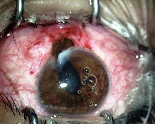
Patients with perforating eye lesions will not be accepted on a commercial flight until the lesion is sufficiently closed, and there is no trapped intraocular air.
Photo: Eye dept.,
Glostrup Hospital
General
The eye is a nearly spherical structure with a diameter of about 22 mm. The eye is a closed compartment with a constant overpressure in the range of 15 to 20 mm Hg. This maintains the shape and function of the eye. Well over 3 ml of fluid is formed in the eye every day, and it drains out primarily through the chamber angle and into the bloodstream. A stop in the drain causes an acute rise in pressure that can easily bring the perfusion pressure in the eye to 0. When that is the case, blood circulation ceases, and acute ischemic damage occurs. The blood pressure in the arteria centralis retinae is about half that measured in the arm.
Open eye injuries often result in loss of pressure and prolapse of intraocular tissue. Typically, there is air in the eye associated with an open eye injury. Likewise, air and medical gas are often used in connection with eye surgery. As the atmospheric pressure decreases in the environment, intraocular air and gas will expand, resulting in an increase in pressure. If the pressure rise is severe, the eye may eventually rupture. In case of open eye injury, further prolapse of intraocular tissue can be seen.
For scheduled and charter flights, air pressure in the cabin will be reduced, which means that flight with patients with air or gas in the eye is contraindicated.
Patients with eye disorders can usually travel unaccompanied if they can see normally with the other eye.
Retinal detachment
The retina is like a membrane in the back of the eye, and the visual function is entirely dependent on its location. If fluid enters the under the retina, the function will decrease in the detached area. This is because the photoreceptors get their oxygen and nutrients from the underlying pigmented epithelium, and when the transport path becomes long, its function diminishes.
The majority of retinal detachments begin in the peripheral field of view, which is why the visual acuity and reading ability is initially unaffected.
A retinal detachment is usually a condition that worsens rapidly with loss of vision if left untreated.
The immediate effort is to stop the progress of the retina detachment. The retina is heavier than the fluid under the retina. This means that the retinal detachment can be stopped or stabilized by backrest. It is recommended that the patient maintains backrest and quiet regimen without reading or movement activity and preferably with eyes closed.
Retinal detachment treatment is operative. In some types of surgery, air, medical gas or silicone oil is injected into the eye.
It is important to distinguish between retinal solutions where
- macula is still attached and where there is still the opportunity to save the central vision.
- macula is detached with loss of central vision.
If the visual acuity is normal, the macula is not detached. If visual acuity is affected, macula is detached to a greater or lesser extent.
If the macula is not detached, emergency surgery is sought. If the macula is detached, the patient should be operated on as soon as possible within 10 days.
If the patient is abroad, surgery should take place if there is a qualified eye surgery center on site.
This is especially true for patients where the macula is not detached. For patients with detached macula, it can be accepted that they travel home for treatment sitting in airplanes as regular passengers.
If there is no qualified eye surgical centre on site, a patient with attached macula should be transported as soon as possible for treatment. Although stretcher transport is the optimal mode of transport, it can be accepted that the patient is placed in business class sitting on a reclining seat
(seat where the backrest can be lowered to 150-170 degrees with extra leg support).
On shorter flights where there is no reclining seat, it may be accepted that the patient is seated in a regular seat. Prior to transport, the patient should have a resting regime as described above. Transport should be as smooth as possible. The patient can be driven seated to and from airports. Seated transport should be arranged at airports. The receiving eye ward must be oriented so that the patient can be received for treatment shortly after returning home.
Air transport of patients after retinal surgery: See Eye surgery.
Glaucoma
Glaucoma is a term for a group of conditions in the eye where there is a progressive loss of nerve fibres in the retina. Often, one of the triggering factors is an increased pressure in the eye.
The most serious manifestation is the acute glaucoma, where the pressure can reach more than 50 mm Hg. Thus, the perfusion pressure becomes 0 mm Hg and there is a clear risk of vision loss. People with high pressure rise in the eye have accompanying pain and affected general condition. They should not be transported but treated locally for reducing intraocular pressure. Once this has happened, the patient can travel by air without restrictions.
Patients with well-treated glaucoma can travel by air without restrictions.
Acute vascular occlusions
There is no acute treatment for either venous or arterial thrombosis in the eye. The condition can be seen in connection with increased pressure in the eye but can also be seen in connection with general inflammatory disease. If possible, the reason should be clarified before returning home. Elevated pressure in the eye should preferably be treated before flight. No special restrictions otherwise.
Ocular injuries
Superficial eye injuries present no problems with air travel.
Open eye damage is an eye trauma with an opening from the inside of the eye to the eye environment. There is an obvious risk of infection, and an open eye injury must therefore be operated and closed as soon as possible at the site of injury. If there is no qualified eye surgery center at the site of injury, the patient should be moved to one. If it is necessary to fly the patient for this primary treatment, it must be performed in an aircraft with cabin pressure of 1 atm. (sea level flight). This is necessary as in the case of open eye trauma there is almost always air in the eye. If the patient is flown under reduced cabin pressure, the air in the eye will expand at the risk of intraocular tissue prolapse.
Air Transport after operation: See Eye surgery.
Eye surgery
In many eye operations, atmospheric air or medical gas is used intraocularly. The eye may be filled with air or gas. An air or gas bubble in the eye will expand with decreasing pressure in the environment, and this may cause an eye-threatening increase in pressure in the eye and ultimately, rupture of the eyeball.
Therefore, if there is air or gas in the eye, air travel with scheduled and charter flights is contraindicated.
After surgery for retinal detachment either with scleral buckling or vitrectomy, there will usually be air or gas in the eye. This also applies after trauma and surgery to such.
Atmospheric air will be gone within a week even with a full eye.
Medical gas in the form of typical C3F8 or SF6 will disappear within 4-7 weeks. However, times may vary from person to person.
Silicone oil is often used as an alternative. An eye filled with silicone oil is no obstacle to air transport.
Air transport of patients after eye surgery:
Ensure that there is no air or gas in the eye. If there is no air or gas in the eye, the patient can travel as a regular passenger.
001. Frontpage
001. Foreword
001. Contributors
001. Aeromedical Problems
012. Planning the Air Transportation of Patients
013. Airline Requirements
015. Transportation of Disabled Persons
016. Cardiac Disorders
019. Gastrointestinal Disorders
010. Central Nervous System Disorders
011. Ear, Nose, and Throat Disorders
012. Eye Disorders
013. Mental Disorders
014. Gynaecological and Pregnancy Problems
015. Transportation of Sick Children
016. Infectious Diseases
017. Orthopaedic Injuries
018. Cancer
120. Acute Mountain and Decompression Sickness
021. Burns and Plastic Surgical Problems
122. Airsickness
123. Jet Lag
124. The STEP System
125. Specialised Transportation of Patients
126. First Aid on Board – Legal Considerations
27. The History of Air Transportation of Patients
28. Oxygen supplementation in flight - a summary
Latest update: 29 - 02 - 2020